Nanosensors for Detecting and Imaging of Cancer Metastasis
This technology introduces a protease-responsive imaging sensor platform for the detection, imaging, and monitoring of tumor development. This platform combines existing protease-cleavable tumor detection sensor with precise tumor imaging optionality by incorporating a pH low insertion peptide (pHLIP) into the sensor scaffold. In preclinical models, active concentration of the sensor at the acidic tumor invasive front by pHLIP produces a demonstrably stronger and more tumor-specific urinary reporter signal than passive tumor accumulation. Because the pHLIP radiolabel is optional, the sensor can be used in a two-step clinical workflow in which all patients receive a non-invasive urine screen and only those with a positive result are exposed to radiotracer for precise tumor localization.
Researchers
-
sensors for detecting and imaging of cancer metastasis
European Patent Convention | Published application -
sensors for detecting and imaging of cancer metastasis
United States of America | Granted | 11,835,522
Figures
Technology
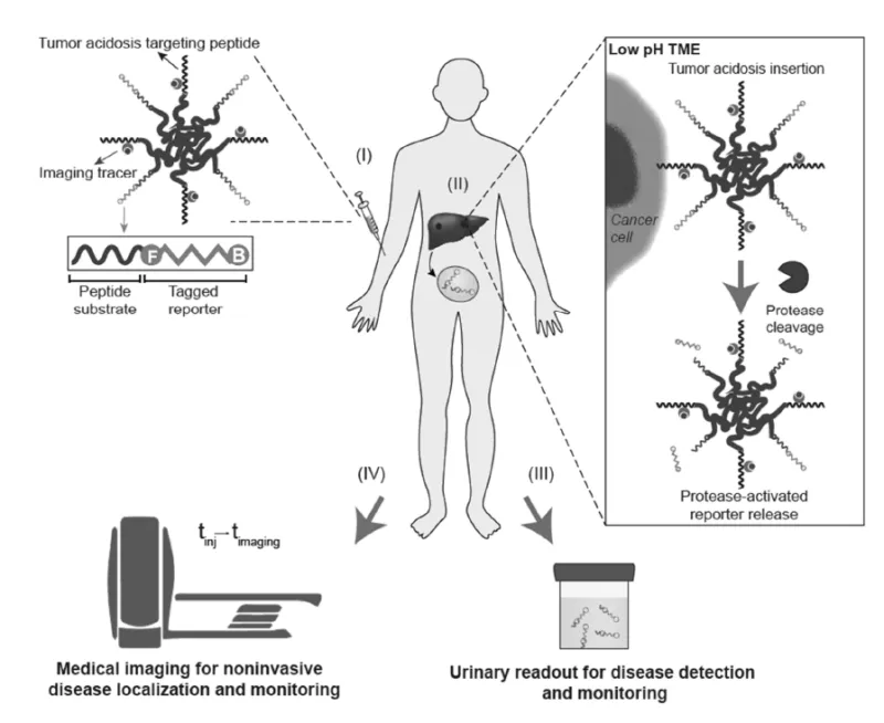
The protease-responsive imaging sensor consists of a modular polyethylene glycol (PEG) scaffold displaying a peptide substrate of Matrix Metalloproteinase-9 (MMP9) or another tumor-associated protease linked to a detectable reporter and a tumor imaging peptide, such as pH low insertion peptide (pHLIP), that can optionally be linked to a radionuclide or other imaging label. Following intravenous administration, the PEG scaffold's size reduces premature renal clearance, enabling continuous circulation and eventual tumor accumulation. Upon encountering the acidic tumor microenvironment, pHLIP undergoes a conformational transition and inserts into tumor cell membranes, concentrating the sensor at the invasive tumor front where MMP9 expression is highest. After MMP9 cleaves the peptide substrate, a small reporter fragment is released that passes through the glomerular filtration barrier, concentrates in urine, and is detectable using methods such as ELISA. When the urinary signal is positive, the same PRISM construct can be loaded on-demand with ⁶⁴Cu for PET-CT imaging to precisely localize the tumor in preclinical models.
Problem Addressed
Metastasis accounts for more than 90% of cancer-related deaths and early diagnosis enables curative surgical intervention for many localized solid tumors, creating a dual imperative for diagnostic tools: non-invasive screening accessible to broad patient populations and highly specific tumor localization for those who screen positive. Current strategies rely on a combination of imaging tests, which suffer from poor specificity in a screening paradigm, and can lead to invasive follow-up procedure and molecular diagnostic assays that, while capable of identifying tumor-associated signals, cannot pinpoint the precise anatomical location of a tumor. By coupling sensitive detection of cancer-associated protease activity with specific tumor localization within a single platform, this technology is designed to provide clinicians with actionable information to guide intervention at multiple stages, from initial diagnosis through treatment monitoring and surveillance for recurrence.
Advantages
- Non-invasive, rapid screening with enhanced tumor-specific sensitivity: The sensor enables non-invasive cancer detection via urine collection with a clinically practical one-hour readout, requiring no specialized collection procedure and amenable to point-of-care implementation. Incorporation of a pH-triggered insertion peptide concentrates the sensor at the acidic tumor invasive front, producing a demonstrably 1.8-fold stronger tumor-specific urinary signal in preclinical models compared to sensors relying on passive tumor accumulation alone.
- Two orthogonal measures of treatment response from a single administration: The urinary reporter reflects tumor invasion capacity, while the imaging signal provides spatially resolved quantification of tumor volume. Together, these readouts can support assessment of tumor presence, metastatic status, tumor progression, and response to therapy.
- Suitability for repeated monitoring: Because the sensor is simple and non-invasive, repeat administration is feasible. Both the urinary and imaging signals scale with tumor burden enabling longitudinal monitoring of tumor progression, remission, stability, or treatment response over time.
- Modular and adaptable pan-cancer platform: The scaffold architecture is designed to accommodate any cancer-associated protease substrate and any compatible radiotracer, positioning PRISM as a broadly applicable platform that can be tailored to specific tumor types, including colorectal cancer.
Publications
Hao, Liangliang, Nazanin Rohani, Renee T. Zhao, et al. “Microenvironment-Triggered Multimodal Precision Diagnostics.” Nature Materials 20, no. 10 (2021): 1440–48.
License this technology
Interested in this technology? Connect with our experienced licensing team to initiate the process.
Sign up for technology updates
Sign up now to receive the latest updates on cutting-edge technologies and innovations.