Intracellular Calcium Sensors for MRI
This invention comprises calcium-sensing probes which can be used in clinical diagnostic imaging with functional MRI to perform neuroimaging, muscular and cardiac imaging, and visualization of immunological activation.
Researchers
-
cell-permeable imaging sensors and uses thereof
United States of America | Granted | 10,933,146
Figures
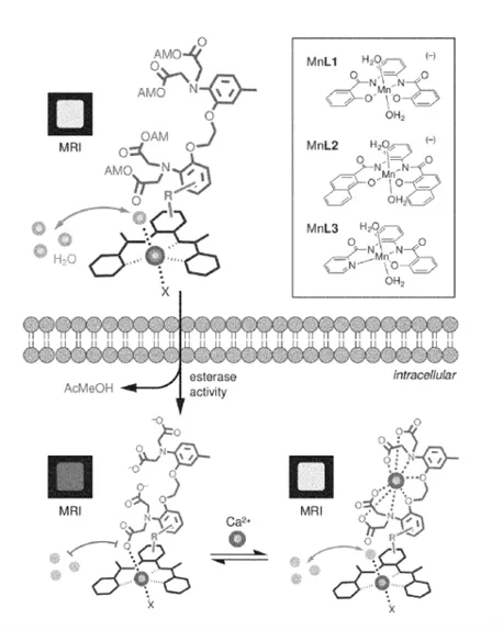
Depiction of cell-permeable calcium sensors for calcium-dependent functional MRI.
Applications
These intracellular calcium sensors are small-molecule complexes comprised of manganese and gadolinium, with planar lipophilic ligands conjugated to the cell-permeable calcium chelator BAPTA. These molecules are able to traverse cellular membranes and enter the cytosol, where they are cleaved and activated by endogenous esterases. Inside the cells, these activated calcium sensors induce changes in T1-weighted MRI contrast upon calcium binding, enabling a noninvasive strategy to visualize calcium signaling within intact tissues for clinical diagnostics.
This technology has widespread applications in the clinic for strategies that require calcium signaling detection, including neuroimaging to identify neuropathies and to plan surgeries, testing of cardiac and skeletal muscle function, and monitoring lymphatic function in inflammation and cancer.
Problem Addressed
Calcium ions are central to signal transduction within cells. They coordinate biological processes ranging from embryonic development to neural function. Being able to detect calcium flux within tissues is essential to the accurate evaluation of cellular activity. However, current methods to measure calcium flux within live tissues for clinical diagnostics remains a challenge. While optical probes for intracellular calcium imaging exist, current strategies to monitor intracellular calcium signaling for clinical applications require invasive surgery to achieve greater tissue penetration. Moreover, attempts to achieve optimal tissue penetration noninvasively forgo image resolution. Magnetic resonance imaging (MRI) is a powerful technique that can provide the basis for deep-tissue calcium imaging in humans and animals. This invention presents a strategy for real-time monitoring of intracellular calcium levels with functional magnetic resonance imaging using novel manganese-based paramagnetic contrast agents which sense calcium.
Advantages
- Non-invasive method to visualize deep-tissue calcium imaging
- High-resolution without sacrificing imaging depth
- Enables the application of functional MRI for visualizing intracellular calcium levels for clinical diagnostics
Publications
Ali Barandov, et al. Sensing Intracellular Calcium Ions Using a Manganese-Based MRI Contrast Agent. Nature communications. 2019 Feb 22. doi: 10.1038/s41467-019-08558-7
License this technology
Interested in this technology? Connect with our experienced licensing team to initiate the process.
Sign up for technology updates
Sign up now to receive the latest updates on cutting-edge technologies and innovations.