Method and Apparatus for Imaging Unsectioned Tissue Specimens
This technology facilitates rapid histological analysis of surgically resected tissues to achieve complete removal of pathological tissues and can be applied in the medical field for intraoperative tissue assessment.
Researchers
-
method and apparatus for imaging unsectioned tissue specimens
United States of America | Granted | 10,416,434 -
method and apparatus for imaging unsectioned tissue specimens
European Patent Convention | Granted | 3,414,553 -
method and apparatus for imaging unsectioned tissue specimens
China | Published application -
method and apparatus for imaging unsectioned tissue specimens
Japan | Granted | 6,961,603 -
method and apparatus for imaging unsectioned tissue specimens
United States of America | Granted | 10,642,019 -
method and apparatus for imaging unsectioned tissue specimens
France | Granted | 3,414,553 -
method and apparatus for imaging unsectioned tissue specimens
Germany | Granted | 3,414,553 -
method and apparatus for imaging unsectioned tissue specimens
United Kingdom | Granted | 3,414,553
Figures
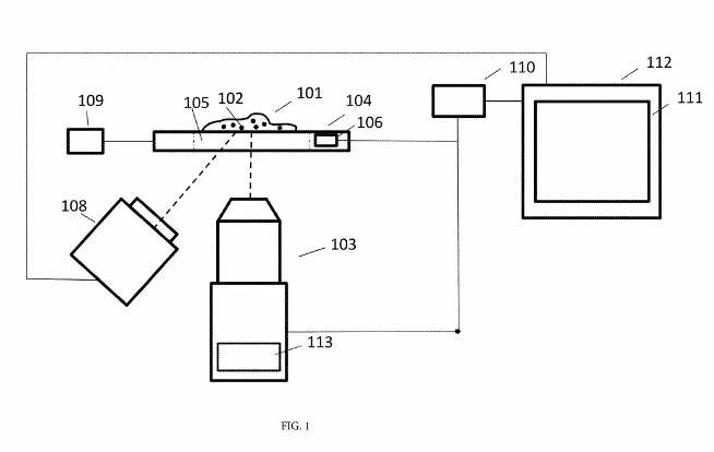
Image 1. Diagram illustrating the optical imaging system, which consists of the primary imaging system, a specimen holder, an auxiliary imaging system, a user input device, a processing unit and a display device.
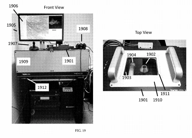
Image 2. Exemplary photographs of the front and top view of the described imaging system including a multiphoton microscope.
Technology
This invention is an imaging system that enables real-time histological analysis of large surgically removed tissues without the need for physical sectioning. The apparatus includes a primary imaging system, auxiliary imaging system, specimen holder, user input device, processing unit, and a user display device. The primary imaging system is an inverted microscope capable of performing optical depth sectioning and configured to acquire a sequence of images. The auxiliary imaging system functions as a camera to acquire auxiliary images covering an area larger than the area of each image acquired by the primary imaging system. The specimen holder positions the tissue of interest on a transparent window with position sensors which measure detect the tissue’s position and translate spatial information to a focal plane in the auxiliary imaging system. The user input device is configured to accept user input which is translated to the specimen holder. The processing unit utilizes the sequences of images acquired by the imaging systems with the spatial information from the specimen position holder to generate a composite representation of the tissue specimen at the cell nuclei resolution. Finally, the display device displays the composite representation of the tissue specimen from the processing unit in real-time.
Collectively, this technology achieves fast histological analysis of large tissue specimens and identifies margins of pathological tissue within the specimen. The system can implement various optical techniques, including multiphoton microscopy and confocal fluorescence microscopy, or techniques based on structured illumination, to detect fluorescent or scattered light at a magnification comparable to a conventional histology microscope, but at an imaging rate suitable for real-time user evaluation.
Problem Addressed
Many surgical procedures, including the resection of solid tumors, require histopathological analysis of the tissue that is removed during the surgery. This process is necessary to determine whether additional therapy or surgery is needed. The resected tissue is preserved with a fixative or by flash freezing, and is then sliced into thin sections for staining and transillumination microscopy to evaluate surgical margins. The physical sectioning of these tissues significantly prolongs the amount of time required to process surgically resected tissues, resulting in this workflow taking at least one day. The tissue assessment is always conducted postoperatively and may necessitate multiple surgeries if the histology indicates that the pathological tissue removal is incomplete.
This technology can mitigate the delays in tissue removal surgery by utilizing an inverted microscope for real-time pathological evaluation of surgically resected tissue, thereby facilitating intraoperative assessment of pathological tissue margins without halting surgical procedures.
Advantages
- Facilitates intraoperative evaluation of surgical margins
- Potential to reduce the number of surgeries to completely remove pathological tissues
- Achieves real-time microscopic image quality comparable to that from conventional histology microscope
Publications
Giacomelli MG, et al. "Multiscale Nonlinear Microscopy and Widefield White Light Imaging Enables Rapid Histological Imaging of Surgical Specimen Margins." Biomedical Optics Express 9, no. 5 (2018): 2457-2475. doi: 10.1364/BOE.9.002457. PMID: 29761001; PMCID: PMC5946802.
License this technology
Interested in this technology? Connect with our experienced licensing team to initiate the process.
Sign up for technology updates
Sign up now to receive the latest updates on cutting-edge technologies and innovations.